
Overview
The Hammer laboratory develops quantitative tools to analyze or mimic biology. We have particular interest in the role of cell adhesion and motility in the immune response. Also, we are interested in developing synthetic tools to mimic biology, such as making artificial cell capsules from synthetic, tunable materials.
Cell Adhesion
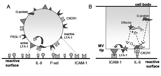
We study how blood borne cells of the microvasculature find their way to targets through molecular zip-coding mediated by cell surface receptors. We have developed novel experimental and theoretical tools to measure and simulate the adhesion of neutrophils – which mediate inflammation – and T-lymphocytes – which regulate the adaptive immune response. We have developed Adhesive Dynamics, a stimulator of adhesion that combines stochastic simulation of receptor dynamics with an accurate mechanical description of a cell. Our diversified suite of algorithms allow simulation of the rolling and stopping of leukocytes. Most recently, we have integrated signal transduction networks within the models, allowing prediction of how defects in signaling cascades affect cell homing (see figure illustrating integrated A. adhesion through multiple receptors and B. signaling networks). Through collaborations both within and outside of Penn, we are testing how molecular manipulations (transfections, siRNA and knock-out mice) affect cell homing in disease.
For an example of a paper in this area, see:
Dooyoung Lee, Jiyeon Kim, Gary A. Koretzky and Daniel A. Hammer, (2012). “Diacylglycerol kinase zeta negatively regulates CXCR4-stimulated T lymphocyte firm arrest to ICAM-1 under shear flow,” Integrative Biology 4:606-614; http://dx.doi.org/10.1039/C2IB00002D
Cell Motility
After leukocytes dock at inflammatory sites, they crawl to their targets. These amoeboid cells crawl quickly but exert small forces. In collaboration with Micah Dembo (Boston University) and Chris Chen (Penn) we have used traction force microscopy (TFM) and micropost arrays, combined with microfluidic gradient chambers, to image the forces that leukocytes exert during chemotaxis and chemokinesis. We have found that neutrophils exert their largest stresses in the rear while dendritic cells exert pulling forces from the front. We are now exploring how the internal molecular machinery of these cells is related to the force generation using knock-out mice and engineered cell lines.
For an example of papers in this area, see:
Brendon G. Ricart, Michael T. Yang, Christopher A. Hunter, Christopher S. Chen, and Daniel A. Hammer, (2011) ”Measuring Traction Forces of Motile Dendritic Cells on Micropost Arrays”, Biophysical Journal; 101(1):2620-2628; http://dx.doi.org/10.1016/j.bpj.2011.09.022
Henry, Steven J., John C. Crocker and D.A. Hammer (2014). “Ligand density elicits a phenotypic switch in human neutrophils,”Integrative Biology 6; 348-356; http://dx.doi.org/10.1039/C3IB40225H
Artificial cells and capsules
We are making artificial cells and drug delivery and imaging capsules from polymers and protein surfactants. Polymersomes are vesicles whose membranes are assembled from block co-polymers. A particularly exciting application has been to make emissive polymersomes, in which porphyrinic molecules synthesized by Michael Therien (Chemistry, Duke) are embedded within the membrane. These vesicles can emit light in the near infrared, are suitable for deep tissue imaging, and with the correct aqueous solute can be engineered to release contents in response to light.
We have also recently made vesicles and other supramolecular assemblies (such as micelles, sheets, and fibers) entirely from protein components, through molecular engineering of naturally occurring surfactant proteins, such as oleosin. These materials allow us to use the tools of molecular biotechnology to make tailored, responsive materials of designed sequence with specific function and responsiveness.
For an example of papers in this area, see:
Kevin B. Vargo, Ranganath Parthasarathy and Daniel A. Hammer, (2012). “Tunable Protein Suprastructures from Recombinant Oleosin”, Proceedings of the National Academy of Sciences USA 109 (29) 11657-11662; http://dx.doi.org/10.1073/pnas.1205426109
Neha P. Kamat, Zhengzheng Liao, Laurel E. Moses, Jeff Rawson, Michael J. Therien, Ivan J. Dmochowski, and Daniel A. Hammer, (2011). “Sensing membrane stress with near IR-emissive porphyrins”, Proceedings of the National Academy of Sciences USA 108 (34) 13984-13989; http://dx.doi.org/10.1073/pnas.1102125108
